
A guide for parents
How blood travels through the heart
01
Follow blood through the heart
The usual blood-flow journey
The usual journey after birth
- Less oxygen
- Mixed blood
- More oxygen
Step 1 of 4
Back to the right side
Blood returns from the body with less oxygen. RA collects it, then it passes into the right ventricle.
How to read this map
Follow the arrows to see connections. This is a map, not the shape or position of a heart. Both sides work together; the story follows one part at a time. A crossing with a white gap is not a connection; a dot marks a junction. With story controls focused, use left/right arrows, Home or End.
Both sides of the heart work together. The steps follow one part of the circulation at a time.
Colours describe this example: blue means less oxygen, red more oxygen, and purple mixing. Blood is always red in real life. Line widths and animation do not measure flow, pressure, oxygen saturation or severity.
The usual journey
- Back to the right side. Blood returns from the body with less oxygen. RA collects it, then it passes into the right ventricle.
- On to the lungs. The right ventricle pumps blood through the pulmonary artery to the lungs, where it picks up oxygen.
- Back to the left side. Oxygen-rich blood returns through the pulmonary veins to LA, then passes into the left ventricle.
- Out to the body. The left ventricle pumps through the aorta to the body. Both sides work together as the journey continues.
Both sides of the heart work together. The steps follow one part of the circulation at a time.
Optional: explore the heart in 3D
One difference at a time
Explore a usual heart
See how the chambers and vessels fit together.
Loading 3D connects to Sketchfab, which receives your IP address and may use cookies. The model may use a lot of mobile data. Sketchfab privacy policy.
Usually
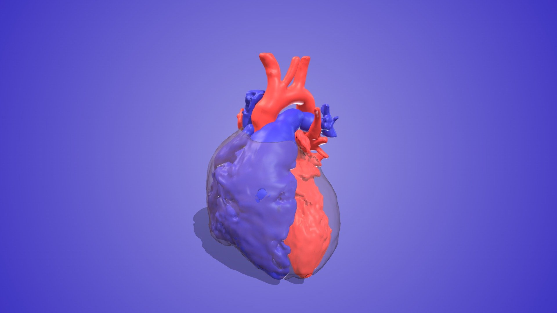
This artery leaves the left ventricle.
Models: 3D Heart Project · CC BY-NC 4.0 · Our labels and explanation await clinical review.
About these pictures and sources
These models show the spaces blood fills inside the heart and its vessels. The typical-heart model also shows a see-through outline of the heart muscle. Colour helps show the parts; blood is never blue.
The labels identify the artery, not its hidden connection to a pump.
Original previews converted to PNG and framed to fit the page, with added labels and captions. The models are from the University of Alberta / Stollery Children’s Hospital. No endorsement is implied.
Sources checked 10 October 2026. Independent clinical review pending.
02
Heart terms and your child’s heart
Doors and walls
The four valves are called tricuspid, pulmonary, mitral and aortic. The septum is the wall between the two sides of the heart. Arteries carry blood away from the heart. Veins bring it back. These names tell you the direction of flow, not how much oxygen the blood carries. Heart differences present from birth can change or narrow the route, or allow blood to mix.
Using a picture together
Ask your child’s doctor or nurse to draw how blood flows through their heart so you can compare the connections. Heartpedia has interactive models you can turn and explore. Ask the team which model matches your child’s heart and what details it leaves out.
03
Questions to ask
Save the questions you want to take to your next appointment. You can edit them in your Journal.
- Could you show us where blood travels differently in our child’s heart compared with the usual route?
- Which valve or blood vessel is affected, and what does that difference mean for how the heart works?
- Could you draw how blood is expected to flow after treatment and explain what will change for our child?
Sources and review
Sources checked 9 October 2026. Independent clinical review pending.
- British Heart Foundation: How your heart works
Accessed 9 October 2026
- GOV.UK / NHS screening: Congenital heart disease: information for parents
Accessed 9 October 2026
- Cincinnati Children’s Hospital Medical Center: Heartpedia mobile app
Accessed 9 October 2026